Intro Block Section Instruction
Form Block Section
Write all of the introduction content below. Only use NON
FULL-WIDTH blocks.
Full-Width blocks can be inserted
outside of this block and will wrap around the form.
Use the custom HTML block below to embed your form.
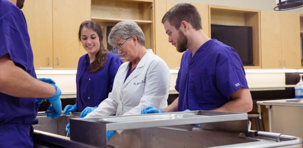
Inspire others with your knowledge of human anatomy.
A firm grasp of human anatomy provides a foundational understanding for healthcare professionals, enabling them to diagnose and treat medical conditions and promote overall wellness. Once you’ve delved deep into the world of the human body, wouldn’t it be great to share those teachings with those pursuing various facets of the healthcare field?
This 12-hour certificate will prepare individuals to teach gross anatomy in a variety of health professions including physical therapy, physician assistants, occupational therapy, speech and language pathology, nursing, medicine, respiratory therapy, radiological sciences, dentistry, optometry, podiatry, dietetics, athletic training, exercise physiology/sport sciences, and chiropractic.
A strong science foundation and a minimum of a Bachelor’s degree is required, as well as a professional and/or graduate degree for admission to the program. PTs, PAs, OTs, SLPs, Nursing, RT, ATC, and Exercise Science College Instructors can all benefit from this certificate to assist in teaching.
The purpose of this certificate is to meet the shortage of qualified professionals to teach anatomy in the health professions. This unique hybrid certificate program includes 4 online courses with a final 4-day intensive course on campus (in May), where human donor dissection leads to increased knowledge, skills and new ways of teaching anatomy and laboratory techniques. Unique to this certificate is the incorporation and application of differential diagnosis and diagnostic imaging utilizing clinical anatomy. Developmental embryology is covered in Neuroscience.
To receive the certificate, one must complete all 12 hours and earn a minimum grade of C in all coursework. Tuition is $699 per credit hour or $8,388 for the full certificate, plus fees. Classes begin Aug. 24, 2026. Certification application deadline is Aug. 14, 2026. In-person lab: May 10-13, 2027; 8:00 a.m. – 6:00 p.m.
Degrees
Why Earn Your Certificate in Clinical Anatomy from HSU?

Why Earn Your Certificate in Clinical Anatomy from HSU?
Online/Hybrid
12-hour Hybrid Certificate Program including online and hands-on human cadaver dissection.
Designed for the Medical Professional
PTs, OTs, SLPs, Nursing, RT, ATC, Exercise Science College Instructors can all benefit from this certificate to assist in teaching.
Outcomes
The purpose of this certificate is to meet the shortage of qualified professionals to teach human anatomy. This certificate will prepare individuals to teach gross anatomy in a variety of health professions including physical therapy, occupational therapy, Physician Assistants, speech and language pathology, nursing, medicine, respiratory therapy, radiological sciences, dentistry, optometry, podiatry, dietetics, athletic training, exercise physiology/sport sciences, and chiropractic.
“As a PT veteran (25+ years) & an amateur anatomy instructor (4+ years), I found this program to be the perfect professional development course. Marsha is awesome and the environment is super conducive to learning.”
Melissa C

Program Details
Click on plus button at the bottom of this Accordion BLOCK (not accordion tab) to create a new tab. Select Accordion TAB block to edit tab headline. Select nested blocks within accordion tab to edit content.
-
Courses
Courses Offered Through 11 Months
- PHYT 7103 Differential Diagnosis and its Anatomical Implication (Online – Spring)
- PHYT 7202 Diagnostic Imaging in Clinical Anatomy ( Online – Summer)
- PHYT 7301 Clinical Anatomy for Anatomy Certification program (Online – Fall)
- PHYT 7304 Clinical Neuroanatomy for Anatomy Certification (Online – Spring)
- PHYT 7308 Clinical Anatomy and Neuroanatomy Lab (Hands on-full cadaver dissection: third week in May)
Clinical Anatomy for the Certification Program PHYT 7301 (3 credits) FALL
Clinical anatomy covers the major structures of the back/ limbs followed by the head/ neck, thorax and abdomen, including pathophysiology, diagnostic imaging, and follows with clinical case scenarios. There is an emphasis on arthrology, histology, osteology, and gross neuromuscular and vascular anatomy of the superficial and deep thoracic and lumbar region, cervical region, upper limb, and lower limb. Emphasis is on general relationships between structures and applied anatomy relevant to the healthcare professions. There is an emphasis on study of the spine, abdominal and pelvic viscera, posterior abdominal wall, thorax, the lungs and heart, neck, head and face, and the cranial cavity and contents. An emphasis on general relationships between structures and applied anatomy relevant to the practice of physical therapy is emphasized as well as to teaching anatomy in health disciplines.Books
1.Gilroy A. MacPherson B. Wikenheiser J. Atlas of Anatomy, General Anatomy and Musculoskeletal System, 4th edition.2020. Thieme Publishers. ISBN-13: 978-1-684-20-203-4.2. Agur AM, Dalley, AF. Moore’s Essential Clinical Anatomy, 7th Edition. 2024.Wolters Kluwer Health. ISBN: 9781975174248
3. Visible Body 4-D4. Other anatomy texts the student may have
Course Objectives: Upon successful completion of this course, the student will be able to:
1. Utilize anatomical terminology and its primary features of human systems.
1.1 Select and utilize anatomical terms correctly orally and in writing.
1.2 Outline the anatomy of the integumentary system.
1.3 Relate the basic anatomy and function of the connective tissues.
1.4 Contrast the basic anatomy and function of muscle and nerve tissue.
1.5 Differentiate the embryological development of the germ layers.
1.6 Compare and contrast the principle features of development of the nerve,
muscle, and skeletal systems.
1.7 Verbalize the histology of structures of the human anatomy.
2. Develop a 3-dimensional image/concept of the skeletal and muscular systems of
the body; with particular attention to the back and limbs.
2.1 Specify the attachments of muscles.
2.2 Relate individual and groups of muscle actions by the line of muscle fiber
from the skeletal attachments.
2.3 Diagram muscle attachments to the skeleton, cadaver and in the living
subject.
3. Verbalize the function of all ligaments through knowledge of specific attachments
and the line of action of ligament fibers.
4. Analyze a 3-dimensional image/concept of the nervous system.
4.1 Differentiate the individual peripheral nerves.
4.2 Determine which nerves go to surrounding structures.
4.3 Integrate muscle innervation to functions.
4.4 Evaluate nerve lesions, at various levels, relate to muscle dysfunction.
4.5 Compare and contrast the cutaneous innervation of dermatomes and
cutaneous innervation patterns as related to the spinal nerve.
4.6 Compare dermatomes, myotomes and sclerotomes of the body
5. Explain the anatomy of the peripheral nervous plexii.
5.1 Diagram, label and relate muscle to nerve and spinal cord segment of
origin of the brachial and lumbosacral plexii.
5.2 Relate spinal cord segments to individual muscle innervations.
5.3 Integrate pre-axial, post-axial determination of innervation and functional
applications
6. Explain and outline the anatomy of the arterio-venous circulatory system.
6.1 Relate the systemic vascular supply to its origin from the heart.
6.2 Select the major vessels to the limbs.
6.3 Integrate anatomical route of vessels relative to regional anatomy,
landmarks and clinical significance of anastomoses.
6.4 Relate the systemic vascular supply to the heart.
6.5 Specify the major vessels of the head, neck, thorax, abdomen and pelvis.
6.6 Integrate anatomical route of vessels relative to regional anatomy,
landmarks and clinical significance of anastomosis
6.7 Relate anatomy of the heart to its blood supply.
6.8 Relate the anatomy of the great vessels at the root of the neck.
6.9 Distinguish the major vasculature of the abdomino-pelvic cavity
6.10 Relate neuro-vascular components of regions of the neck and face
7.0 Explain embryology of the heart and relate this development to possible
congenital abnormalities
8.0 Verbalize the anatomy of the abdomino-pelvic cavity.
8.1 Outline the muscles and their innervation of the anterolateral abdominal
wall, respiratory and pelvic diaphragms, and the posterior abdominal wall.
8.2 Compare and contrast the relationships and orientation of the
abdominal and pelvic viscera.
8.3 Relate concepts of retro-and intra-peritoneal viscera and the mesentery.
9.0 Diagram the anatomy of the head and neck.
9.1 Differentiate the major bones, foramina and landmarks of the skull.
9.2 Contrast muscles of the neck, and those of facial expressions and
mastication.
10.0 Investigate anatomy to clinical scenarios
10.1 Explain dysfunction or abnormalities that occur with an injury to a muscle or
nerveClinical Neuroanatomy for Anatomy Certification PHYT 7304 (3 credits) SPRING
Clinical Neuroanatomy covers information related to function of the human peripheral and central nervous system. Particular attention is given to neuroscience, neuro-embryology, sectional anatomy of the brain, brainstem, spinal cord and the related neural pathways for the motor and somatosensory systems. Emphasis is on general relationships between structures and applied anatomy relevant to the healthcare professions. Clinical case scenarios and various teaching methods will be incorporated into this class.Course Objectives: Upon successful completion of this course, the student will be able to:
1. Relate anatomical terminology of the nervous system, communicating proper use of terms in a clinical context.
2. Develop a 3-dimensional image/concept of the nervous system, contrasting the anatomy of the brain, brainstem and spinal cord.
3. Explain spinal cord anatomy, including describing the general organization of gray and white matter as well as the concept of long tract systems of the spinal cord, and discussing the sensory and motor pathways.
4. Relate selected lesions of the nervous system to clinical signs and symptoms by discussing the results of lesions to selected areas of the brain, brainstem, and spinal cord.
5. Explain the basic anatomy and circuitry of the cerebellum, describing the connections of efferent and afferent connections as related to the results of cerebellar lesion clinical signs and symptoms.
6. Discuss the basal ganglia and the extrapyramidal system, describing the anatomical substrate and functional aspect of the systems and relating the results of clinical lesion signs and symptoms.
7. Explain the neural substrates for pain, discussing the sensory receptors, sensory pathways and termination for pain perception related to the neural connections and clinical aspects of mechanisms for pain control.Books:
1. Neuroscience 6th Edition (2022) by Laurie Lundy-Ekman PhD PT, Publisher: Saunders; 6th edition (June 15, 2022) (ISBN-13: 978-0323792677)
2. 7th edition of Goodman and Snyder Differential Diagnosis for Physical Therapists (ISBN -13: 978-0323722049)
3. various online neuroscience and/or neuroanatomy resourcesOPTIONL Available through McGraw Hill/FA Davis Access PT online offered through HSU
Differential Diagnosis and Its Anatomical Implication PHYT 7103 (1 Credit) SPRING
This course will utilize basic concepts taught in anatomy to assist the learner in differentiating and identifying cutaneous, somatic, visceral, neurogenic, referred and low back pain by identifying red flags, and examining systemic illness.Course Objectives: Upon successful completion of this course, the student will be able to:
1. Differentiate the types of serious sources of low back pain.
2. Visualize the process and the clinical uses of several heuristics for differential screening.
3. Explain the types of patient data that falls under the category of symptom investigation, including red flag information requiring physician contact and risk factors that require monitoring during patient management.
4. Value the role and responsibilities of differential diagnosis in patient management.
5. Hypothesize signs and symptoms associated with medical disorders that may result in patient pain syndromes.
6. Evaluate data from examination to make clinical judgments in the diagnosis and management of the patient.
7. Build useful knowledge needed for comprehensive history taking and physical examination for differentiation of chest and abdominal pain disorders.
8. Differentiate when abdominal pain or chest pain is musculoskeletal versus visceral in nature.
9. Evaluate risk factors relevant to medical screening and for monitoring and modifying in the screening process.
10. Determine constitutional symptoms that suggest systemic illness.
11. Conduct a systems’ review and frame questions to obtain maximal patient cooperation in the process.
12. Differentiate vascular versus neurogenic back pain.
13. Formulate the key areas of questioning/information gathering required for assessment of patient’s pain.
14. Justify the most likely source of pain (i.e., cutaneous, somatic, visceral, neurogenic, or referred), given a description of a patient’s pain.
15. Construct a body diagram of referred pain and hypotheses as to the sources of the pain.
16. Validate signs and symptoms of psychological disorders that may affect a patient’s response to pain and the tools used in this process.
17. Assemble & Utilize knowledge of regional screening conditions and useful tests to differentiate atypical conditions from typical musculoskeletal conditions.
18. Illustrate a neurological screening exam to detect limb pain and neuropathy, to include cranial nerves and peripheral nerve integrity.
19. Simulate an upper and lower quarter screening examination.
20. Differentiate serious red flags that require immediate physician referral and consultation.
21. Develop effective approaches to hip/pelvic/groin, neck/back, shoulder/upper back, and chest/breast screenings based on the work of Goodman and Heick..Book: Goodman and Snyder’s Differential Diagnosis for Physical Therapists :Screening for Referral. 2022; 7th edition. Elsevier. Paperback ISBN: 9780323722049; eBook ISBN: 9780323833691
Diagnostic Imaging in Clinical Anatomy PHYT 7202 (2 credits): SUMMER
Course Description: Diagnostic Imaging will be taught online through use of different radiological images, including Clinical diagnostic imaging includes radiology, MRI, diagnostic ultrasound, CT scan, etc.Course objectives: By the end of the course, the student will be able to:
1. Appraise multiple forms of diagnostic imaging (Radiographs, CT scans, Diagnostic Ultrasound, MRI, etc):
1.1. Position plain radiographs or other diagnostic images correctly for viewing.
1.2. Illustrate key anatomical landmarks and structures of the human body relative to systems, location, and planes of the body through radiographic imaging.
1.3. Formulate an enhanced working vocabulary of diagnostic and musculoskeletal imaging terminology.
1.4. Compare and contrast normal and abnormal features on the radiograph/diagnostic images, including, but not limited to bony abnormalities and fractures.
1.5. Analyze the radiograph/image in terms of view and anatomical area being studied.
1.6. Distinguish signs, symptoms, and imaging appearances of a broad spectrum of musculoskeletal and non-musculoskeletal pathology.
1.7. Assess the validity of various forms of imaging based on evidence-based criteria.
2. Utilize the information to be gained using the various imaging techniques.
2.1. Examine the specific purposes for using different imaging techniques, including radiographs, MRI, CT scan, Diagnostic Ultrasound, and bone scans.
2.2. Compare and contrast the advantages and disadvantages of each technique.
2.3. Formulate factors considered to determine the correct choice of imaging techniques.
2.4 Engage in the diagnostic process to establish differential diagnosis utilizing imaging and determine its relevance to clinical decision-making.
2.5 Mentor peers on musculoskeletal imaging according to the principles of evidence-based practice.
2.6 Utilize imaging to enhance differential diagnosis and clinical decision-making plan
2.7 Formulate concerns of diagnostic imaging as related to diagnosis and health of the individual.
2.8 Investigate a Dexascan and its evaluation of skeletal integrityBOOKS:
McKinnis, LN. Fundamentals of Musculoskeletal Imaging, 5th edition. (2020). FA Davis. ISBN-13: 9780803676022 (available online through HSU)Clinical Anatomy and Neuroanatomy Laboratory PHYT 7308 (3 credit) MAY
This course is the culmination of the certificate program and incorporates a 5-day, onsite, hands-on lab component to include cadaveric dissection, regulations of the anatomy lab and teaching ideas.
I. Clinical Anatomy Laboratory (28 hrs-2 credit)
Objectives: By the end of the course, the student should be able to:
1. Analyze the anatomical features of articular cartilage, bone, ligaments, muscle,
tendons and neural tissue in the upper extremity, lower extremity and spine.
4. Examine the musculoskeletal anatomical variations between individuals and its implications on clinical practice.
5. Correlate surgical conditions utilizing cadaveric dissection to clinical practice.
6. Integrate cadaveric dissections of the heart, lungs, diaphragm, and GI tract into clinical case scenarios, differential diagnosis and treatment.
7. Evaluate the effects of aging and selected disease processes on musculoskeletal tissues.
8. Investigate specific insertions, actions, nerve supply and vascular supply of muscles of the upper extremity, lower extremity and spine and it effect on movement.
9. Verbalize lab preparation/ handling of specimens and the requirements for implementing/sustaining a human anatomy cadaver laboratory.
10. Develop and perform dissection skills appropriate for teaching cadaveric
dissection combined with utilization of diagnostic images/diagnostic ultrasound/and dry needling application.
11. Construct and perform a teaching project to incorporate evidence-based
anatomical knowledge and cadaveric dissection.
12. Contrast and compare anatomical findings with differential diagnosis utilizing
dissection of organs with evidence-based practice.II. Neuroanatomy Laboratory (20 hr-1 Credit)
1. Actively teach anatomical structures of the neuro system to other students
2. Perform Dissection of human cadaver combined with diagnostic images of areas of the brain and spinal cordBOOKS: Previous anatomy books used in PHYT 7301 and PHYT 7304 will be used.
-
Requirements
A strong science foundation and a minimum of a Bachelor’s degree is required, as well as a professional and/or graduate degree for admission to the program.
- 1 letter of recommendation – from a supervisor or academic professor. Must be on letterhead, dated and signed with professor credentials
- Resume/CV
- Personal Statement – explaining why you want to complete this program (Dated, and signed with your credentials)
- Transcripts from all universities/colleges
- Background Check (choose the Non-Employee Background option)
- Ethics Statement
-
Tuition & Fees
Tuition Gen Fee Online/Lib Other Total Fall 2025 2,097.00 253.00 100.00 2,450.00 Spring 2026 2,796.00 253.00 200.00 3,249.00 Summer 2026 3,495.00 305.00 100.00 200.00 4,100.00 Totals: 8,388.00 811.00 400.00 200.00 9,799.00 Tuition/HR: 699.00 -
Gould Anatomy Faculty

Dr. Marsha Rutland PT, ScD.
Professor of Physical Therapy- Shelton-Lacewell Endowed Scholar
- B.S., Physical Therapy, UT Southwestern-Dallas, 1979
- M.Ed., Hardin-Simmons University, 2002
- Sc.D., Orthopedics, Texas Tech Health Science Center, 2008
- Previous Board Certified Clinical Specialist in Orthopedic Physical Therapy (2003-2023)
- Certified Orthopedic Manual Therapist (COMT-IAOM)
- Certified Dry Needler (IC-DN- IAOM)
- Certified Strength and Conditioning Specialist (CSCS)

Wendy K Anemaet
Professor of Physical Therapy
Education:- B.S. Biology, B.S. Pre-Med, B.S. Athletic Training, Mount Vernon Nazarene University
- MS, Physical Therapy, The University of Southern California
- PhD, Aging Studies, University of South Florida
- DPT, Physical Therapy, Regis University
-
Student Testimonials
“Even as someone who has taught anatomy for a few years, I thoroughly enjoyed this program and learned a ton! Feel free to contact me” kpellow@apu.edu
“As someone without a clinical background and coming purely from a STEM-focused education, I was initially intimidated by the clinical depth of the anatomy certification program. After speaking w/ Dr. Rutland, I applied and it turned out to be the perfect course to expand my clinical anatomy knowledge.” – Robert W. – 2025
“This program is well worth your time and effort. Excellent instruction, excellent feedback, excellent collaboration. Dr. Rutland is an absolute treasure to this program and to our profession.” – Kendra W. kmcreynolds@emoryhenry.edu – 2025
“The 1-week anatomy lab was very helpful. My learning gave me alternate ways / techniques to find structures that are typically difficult to find.” – 2025
“This program has been invaluable in developing my teaching and dissection skills I am now able to connect anatomy more deeply to our entire curriculum.” – Kristina D. – 2025
“This is a wonderful course whether you are in the beginning stages of designing a course, teaching for the first time, or if you have 20+ years of anatomy teaching experience. The didactic information is useful to anyone at whatever stage you are in and not only contains a wealth of information but a treasure trove of teaching tips and ideas that even the most seasoned anatomy instructor can learn from and use. The week of dissection is an amazing experience and interacting with colleagues from multiple disciplines exposes you to a plethora of ideas from dissection techniques and tips and you are given the freedom to explore as you wish.” P-NC, Class of 2024
“As a new instructor of a DPT anatomy course, this program gave me so many ideas to use in my teaching to enhance student learning and make learning anatomy fun, while also increasing my confidence to teach this content. The set-up of this certificate program encourages a lot of sharing of ideas that really help new or seasoned instructors of anatomy. The dissection week at HSU was so helpful to learn from the instructors at HSU and also classmates related to unique dissection techniques and experiences. Dr. Rutland does a fantastic job of facilitating discussion and learning and makes you think outside of the box for creative ways to teach anatomy.” -Kyle C, Class of 2024
“This was a wonderful educational experience designed for the working professional. It included lots of helpful information in a flexible format which built up to an in-person experience in the lab.” -Melodie K, Class of 2023
“I am so blessed to have been a part of this anatomy certificate program. I am walking away with an immense amount of knowledge and a refreshed love for anatomy.” -Tiffany F, Class of 2023
“The Gould Graduate Certificate in Human Anatomy at Hardin-Simmons University has been a transformative program for me as a young faculty member. I have gained a trove of creative and effective methods for teaching anatomy to my students, and I have gained confidence in my ability to lead my students through dissection. Dr. Rutland and her team are incredible educators, humble, deeply knowledgeable, and very encouraging and thoughtful.” -Hugo K, Class of 2023
“I have been using my newfound knowledge that I learned every week! Looking back, I found the program to be very helpful.” -J.S., Class of 2021
“Great way for non-traditional PhD anatomists to expand anatomy skills.”
“I highly recommend the Anatomy Certification Program to clinicians and academics alike. You will be intellectually stimulated and stretched in a welcoming, non-punitive environment.” – Ken W.
“The HSU Anatomy Certification Program helped me to get into the frame of mind of the teacher, to help take what I knew of anatomy and present it to a learner in a way that was beneficial to both teacher and student. It has helped me to adjust my teaching to help increase student retention and clinical application.” – Seth M., ’21
“I’ve already used at least five teaching methods that I learned in the anatomy course taught by Dr. Rutland and it’s not even the end of week three of class. I feel a lot more confident in reviewing radiographs with students thanks to the diagnostic imaging course as well. This program was invaluable is providing me with teaching ideas that my students can remember and apply clinically in a more meaningful way. Connecting with and learning from other professionals, PT, PA, and MD was also an amazing experience that I otherwise would not have had outside of this program.” – Lara D., ’21
“The certification program is highly-informative and user-friendly. The program is very challenging and I found it to be a great way to enhance and fortify my anatomy knowledge. The faculty are extremely knowledgeable, caring, and supportive. All of the classes were indeed helpful; however, the dissection class was by far one of the most educational experiences I could have hoped for. This program, without a doubt, was well worth every minute of my time.” – Darryn W., ’21
“The Certification in Clinical Anatomy at HSU was a great experience and I am very happy to have taken it. As an anatomy professor in a DPT program without a PhD degree in anatomy (and no current desire/time to go that route), taking this course was a fantastic option. Major components of musculoskeletal anatomy and neuroanatomy felt like the major themes I am coming away from the course with, but inclusion of diagnostic imaging and differential diagnosis also seemed to round out the certificate program well. The faculty are extremely passionate about helping us improve our knowledge base to impact our students well. The program definitely felt geared towards PTs in academia but there were others in the class who clearly gained a lot from the program. I’ve implemented many things into my courses that I learned during this program. Would highly recommend!” – AJP, ’21